Wrist Xray Interpretation OSCE Guide Geeky Medics
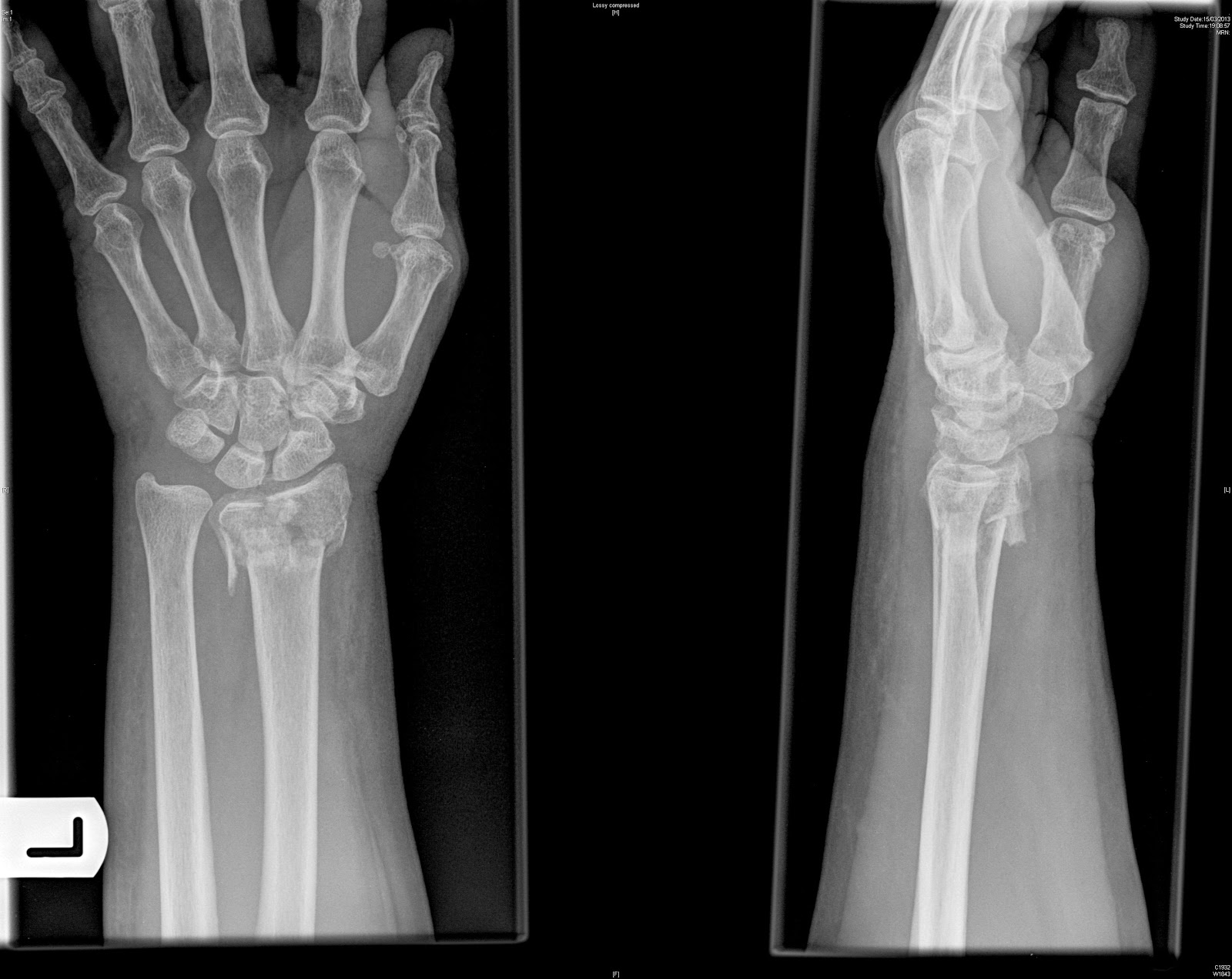
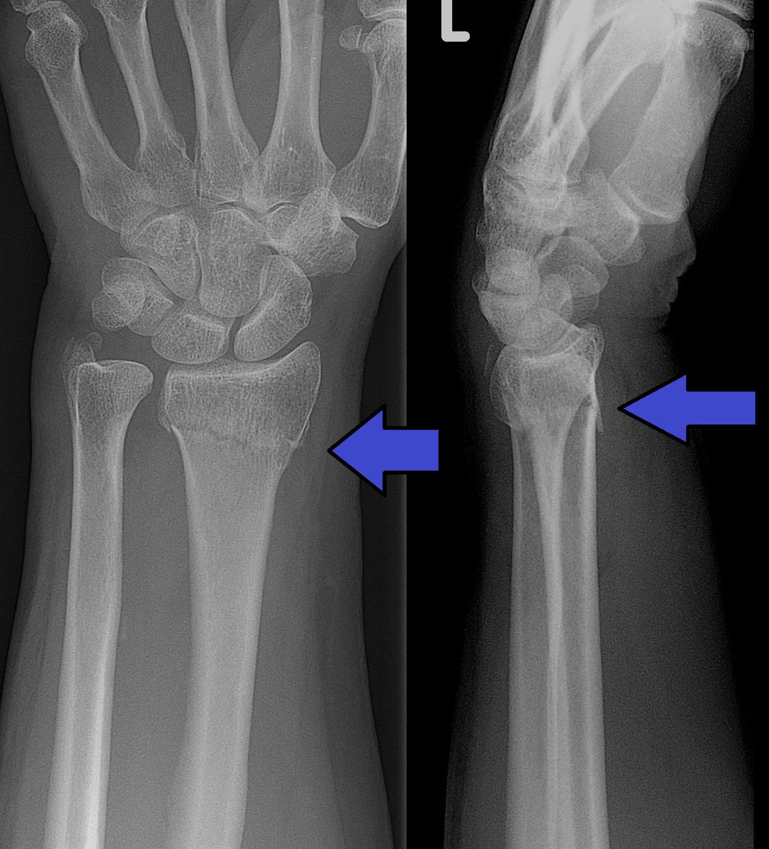
Age: 35 years Gender: Female Radiographs of wrist x-ray Frontal Lateral There are intra-articular fractures of the right distal radius and ulna with significant shortening and volar angulation of the distal fracture fragments. 1 case question available Case Discussion

Κάταγμα Κερκίδας Τραύμα Ευστράτιος Καβρουδάκης, Χειρουργός,Ορθοπαιδικός,Τραυματιολόγος
Introduction Wrist trauma is a common presentation to the emergency department and X-ray is typically the first-line investigation used to identify bony injuries. This guide provides a step-by-step approach to interpreting wrist X-rays and includes examples of the key pathology you may come across. Anatomy

Smith fracture, Xray Stock Image F034/8251 Science Photo Library
Smith fracture - mechanism of injury, X-ray, treatment Smith fracture is a fracture of the distal radius. Although it can also be caused by a dire Show more Show more 1y ago ORTHOfilms

Colle`s and Smith`s fracture YouTube
Barton's Fracture. This is an intra-articular fracture of the distal radius with associated dislocation of the radio-carpal joint. A Barton fracture can be described as volar (more common) or dorsal (less common), depending on whether the volar or dorsal rim of the radius is involved. Figure 2 - Schematic demonstrating difference in.
Smith fracture, Xray Stock Photo Alamy
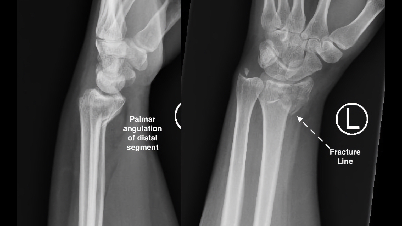
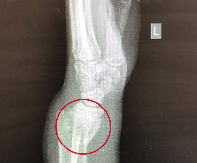
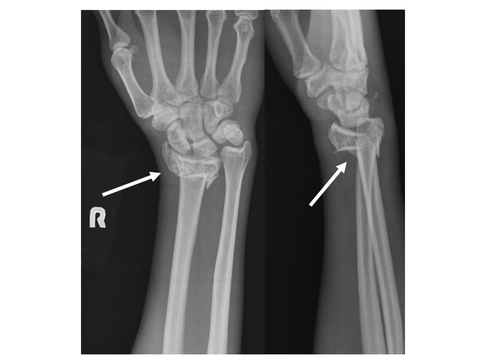
Presentation History of fall on a flexed wrist Patient Data Age: Adult Gender: Male x-ray Impacted fracture of the distal radius with palmar angulation and evidence of intra-articular involvement. There is also ulnar styloid fracture. Case Discussion The images represent Smith fracture 2 articles feature images from this case
The Wrist
This injury produces what is known as a "garden-spade" deformity on X-Ray. Colles' and Smith's fractures often occur in isolation but can have other associated injuries. Isolated radial shaft fractures can occur at any location along the bone. The mechanism of injury for isolated distal third radial shaft fractures is similar to Smith.
Fracture Treatment Osteoporotic Fracture
OBJECTIVE. Fractures of the distal radius are common and frequently encountered by the radiologist. We review the epidemiology, classification, as well as the concept of instabil-ity. Salient qualitative and quantitative features of the distal radius fracture identifiable on the routine radiography series are highlighted.
Explaining What Smith's Fracture Is Physioroom Blog
A short lecture on the X-ray findings in Smith's fracture with clear X-ray illustrations.

Common Distal Radial Fractures UW Emergency Radiology
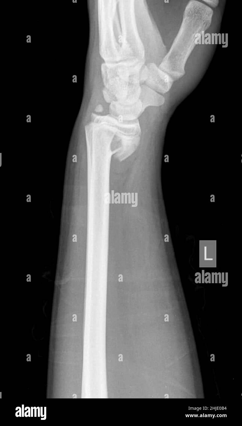
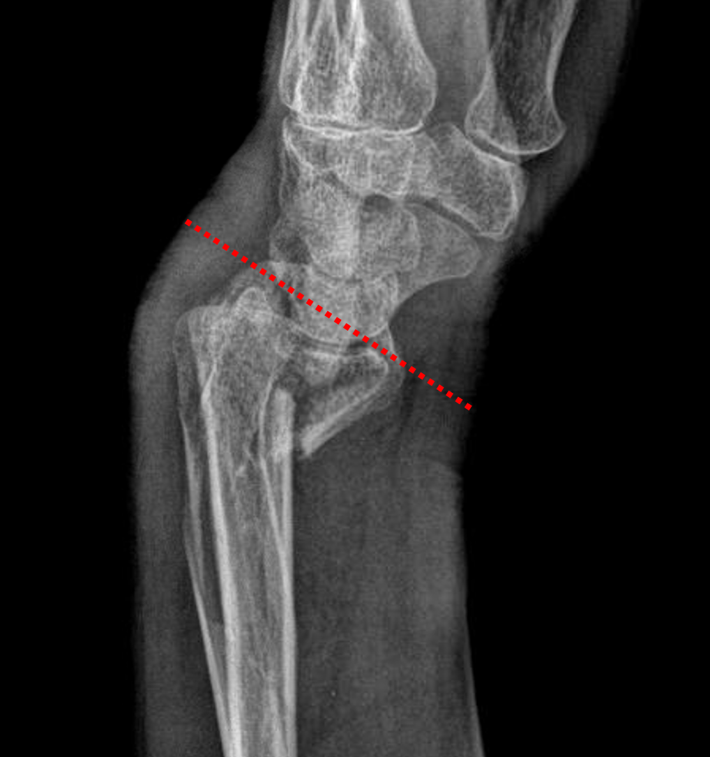
Age: 40 years Gender: Male x-ray Frontal Lateral There is an impacted extra-articular transverse fracture of the distal radius with palmar angulation. Case Discussion The images represent Smith fracture, classically an extra-articular transverse fracture and can be thought of as a reverse Colles fracture. 1 article features images from this case
Smith Fracture (Distal Radius Fracture) Definition & Treatment
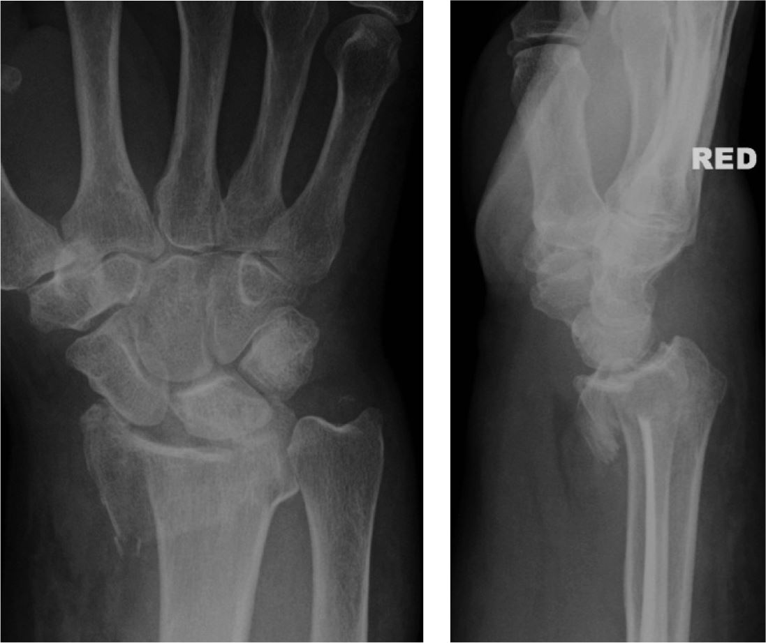
Age: 50 years Gender: Male x-ray Frontal Lateral Lateral, frontal X-ray wrist joint AP and lateral views showing a fracture involving the distal radius with volar angulation of the fractured distal fragment, representing a Smith fracture. Widening of the scapholunate interval is also noted. 2 case questions available Case Discussion
Smith fracture (Frykman IV)
Smith fractures usually occur in one of two ways: a fall onto a flexed wrist direct blow to the back of the wrist Radiographic features The fracture can be split into three types, although in practice a description suffices 1,2: type I extra-articular transverse fracture through the distal radius most common: ~85% type II
Trauma Xray Upper limb gallery 2 Colles' fracture
This fracture is often distinguished from the Colles and Smith fractures by the presence of intraarticular radiocarpal joint involvement [8, 9] . Fig. 3A —Volar and dorsal Barton fractures. A, Frontal ( A ) and lateral ( B ) radiographs in 44-year-old man with volar Barton fracture after motorcycle crash and frontal ( C ) and lateral ( D.

Smith Fracture ATL Physio
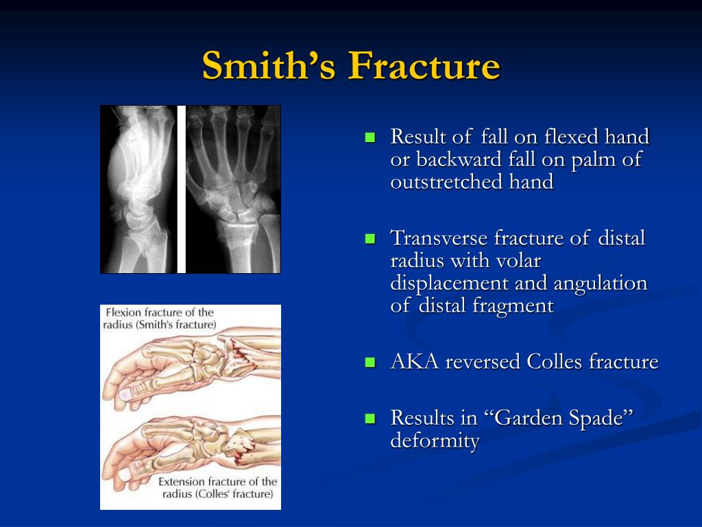
A Smith's fracture, is a fracture of the distal radius. [1] Although it can also be caused by a direct blow to the dorsal forearm [2] or by a fall with the wrist flexed, the most common mechanism of injury for Smith's fracture occurs in a palmar fall with the wrist joint slightly dorsiflexed. [3]

Smith Fracture
Forearm fracture/dislocation. The radius and ulna form an anatomical unit, joined throughout their length by an interosseous ligament and stabilised at the elbow and wrist, thus forming a ring. If there is a fracture of the shaft of one of these bones with visible shortening, there will likely be dislocation at the wrist or elbow of the other.
Chapter 2 Dr Vivek Pandey
There are three types of Smith's fractures. Type I is the most common with a transverse break outside the wrist joint. Type II, also called a reverse Barton fracture, is an intra-articular fracture, or a fracture that occurs on the articular surface (i.e., joint surface) of the radial-ulnar joint.
Dinner Fork Vs Garden Spade Deformity Fasci Garden
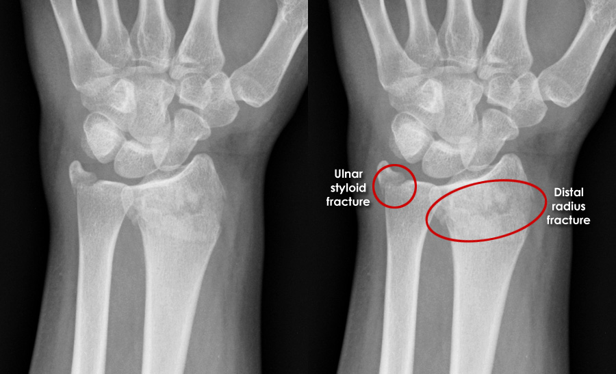
Smith's fracture. Smith's fractures occur in younger patients and are the result of high energy trauma on the volar flexed wrist. Volar comminution and intraarticular extension are more common. On the left an extraarticular Smith's fracture with palmar and radial angulation and displacement. There is also an avulsion of the ulnar styloid process.