The Radiology Assistant Cerebral Venous Thrombosis
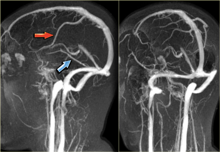
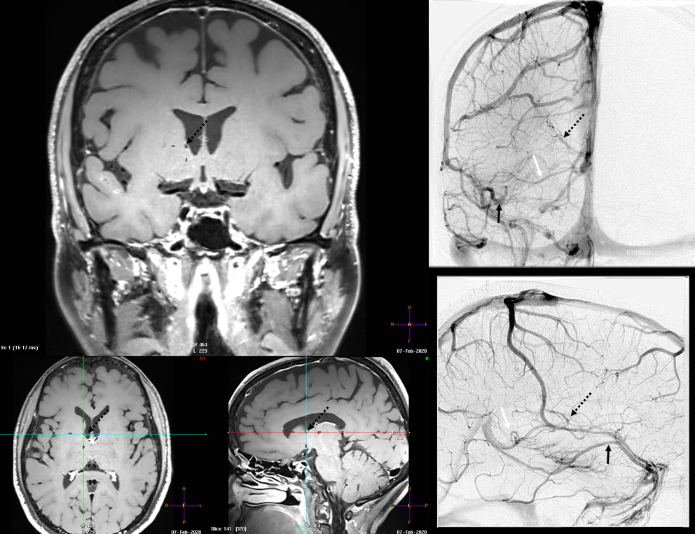
MR venography sequences allow for an initial positive diagnosis of cerebral venous thrombosis and also for monitoring the thrombus and visualizing its partial or complete recanalization. Complete recanalization is not necessary for symptom improvement.

Cerebral veins Image
MRI or MRI venography (MRV) are powerful techniques, provided the radiologist is aware of critical diagnostic pitfalls. In selected cases, cerebral digital subtraction angiography (DSA) can facilitate both diagnosis and anticoagulant/transcatheter thrombolytic therapy improving clinical outcome.

Cerebral vein thrombosis internal Image
Perinatal venous infarcts are underrecognized clinically and at imaging. Neonates may be susceptible to venous infarcts because of hypercoagulable state, compressibility of the dural sinuses and superficial veins due to patent sutures, immature cerebral venous drainage pathways, and drastic physiologic changes of the brain circulation in the perinatal period. About 43% of cases of pediatric.
Intracranial Venous System Overview Radiology Key
The range of intracranial venous anomalies in children differs from that in adults. As a commonly encountered highly morbid disease, sinovenous thrombosis has been discussed extensively in the literature, and the associated imaging considerations are similar in pediatric and adult patients. The authors shift the focus to less frequently discussed cerebral venous diseases in pediatric patients.

Variations of the Superficial Middle Cerebral Vein Classification Using Threedimensional CT
The cerebral venous system comprises superficial and deep veins, which contain nearly 70% of the brain's blood volume and play a crucial role in maintaining normal cerebral perfusion.

Lateral superfical veins of the brain Image
Advanced Imaging Equipment. Long Beach Medical Center utilizes a 320-slice computed tomography (CT) scanner that provides clear images of the brain to diagnose areas affected in a matter of minutes rather than hours and determine the best course of treatment. Conditions. Arteriovenous malformations (AVM) Cerebral aneurysm
Superficial Cerebral Veins Radiology Key
CVT is difficult to diagnose clinically because patients can present with a wide spectrum of nonspecific manifestations, the most common of which are headache in 89%-91%, focal deficits in 52%-68%, and seizures in 39%-44% of patients. Consequently, imaging is fundamental to its diagnosis.
The Radiology Assistant Cerebral Venous Thrombosis
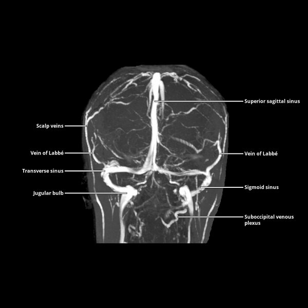
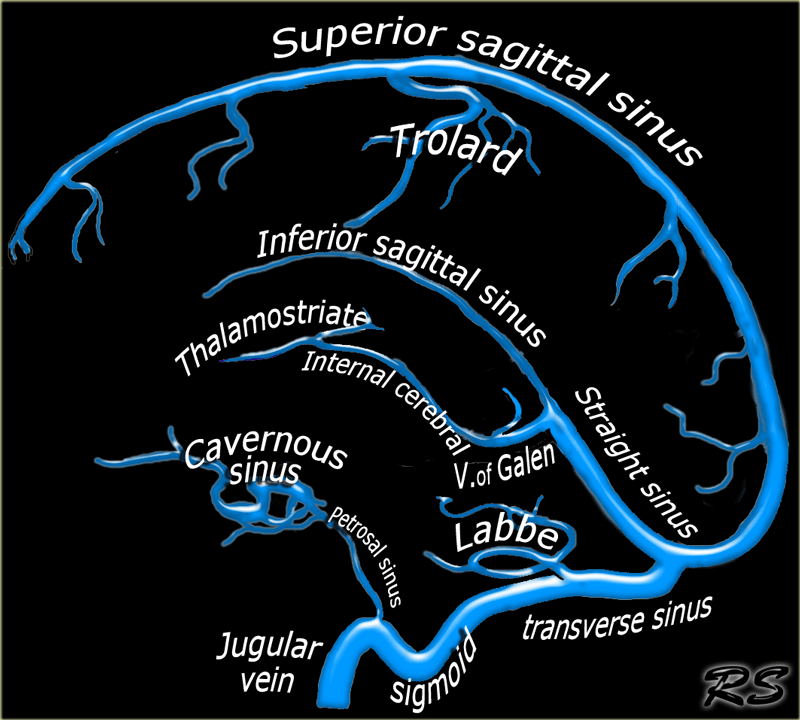
The cerebral veins drain the brain parenchyma and are located in the subarachnoid space. They pierce the meninges and drain further into the cranial venous sinuses. The cerebral veins lack muscular tissue and valves. The cerebral venous system can be divided into: superficial (cortical) cerebral veins deep (subependymal) cerebral veins

Cerebral Venous Thrombosis Radiology Key
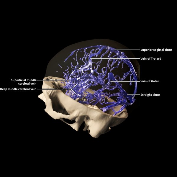
The internal cerebral veins unite with the basal veins (of Rosenthal) to form the great cerebral vein (of Galen) just beneath the splenium of the corpus callosum in the quadrigeminal cistern. The confluence of the great cerebral vein and inferior sagittal sinus forms the straight sinus.
The Radiology Assistant Cerebral Venous Thrombosis
The purpose of this article is to review the clinical presentation and basic pathophysiology of the disease; review the approach for radiologic investigation, including emerging technology such as CT venography; review the imaging features of CVT; and show common pitfalls associated with the radiologic evaluation of this diagnosis.

Cerebral Venous Anatomy. Radiology, Medical anatomy, Anatomy
In recent years, imaging technology has allowed the visualization of intracranial and extracranial vascular systems. However, compared with the cerebral arterial system, the relative lack of image information, individual differences in the anatomy of the cerebral veins and venous sinuses, and several unique structures often cause neurologists and radiologists to miss or over-diagnose. This.

Atypical Deep Cerebral Vein Thrombosis with Hemorrhagic Venous Infarction in a Patient Positive
About Cerebral venous system Last revised by Mendel Castle on 19 May 2018 Edit article Citation, DOI, disclosures and article data The cerebral venous system, somewhat unlike the majority of the rest of the body, does not even remotely follow the cerebral arterial system.

MRI Brain Vascular Anatomy Mri Scan Images Mri brain, Mri, Thrombosis
We report the development of a head-mounted photoacoustic fiberscope for cerebral imaging in a freely behaving mouse. The 4.5-gram imaging probe has a 9-µm lateral resolution and 0.2-Hz frame.
Internal Cerebral Vein
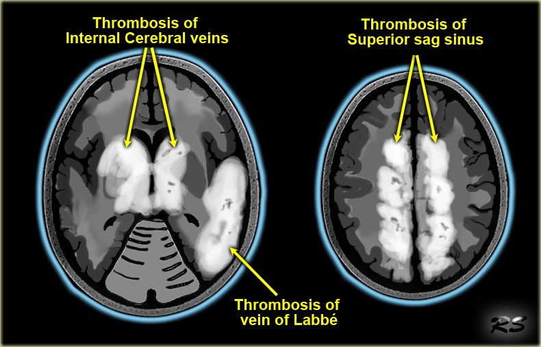
Cerebral venous thrombosis (CVT) is defined as the presence of a thrombus within a venous sinus, superficial intracranial vein, or deep intracranial vein. It is an uncommon condition that is potentially reversible if diagnosed and treated appropriately and promptly.
Presentation1.pptx, radiological imaging of cerebral venous thrombosi…
The deep cerebral veins drain the deep white matter and grey matter that surround the basal cisterns and ventricular system. The deep veins are responsible for the outflow of approximately the inner 80% of the hemisphere.

Fig 1. Multisection CT Venography of the Dural Sinuses and Cerebral Veins by Using Matched
The great cerebral vein , also known as the vein of Galen or great vein of Galen, is a short valveless midline venous trunk that drains the deep parts of the cerebrum, brainstem and parts of the posterior cranial fossa. Gross anatomy